SemiCOL Challenge
SemiCOL Challenge is a computational challenge to one of the most common human malignant epithelial tumors – colorectal cancer.
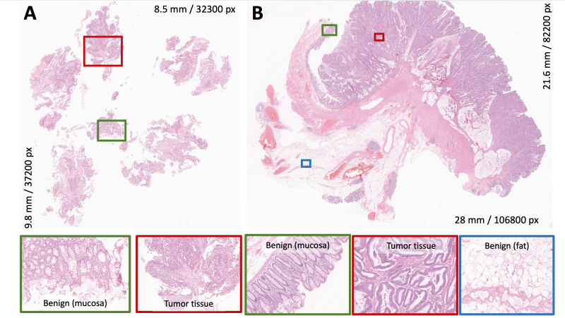
Tumor tissue detection in the pathological material is one of the routine diagnostic tasks for pathologists. There are two types of specimens being submitted to pathology departments (Figure 1):
- 1. Biopsy (small pieces of tissue sampled during endoscopic procedure from suspicious regions; Aim: confirmation or rejection of tumor diagnosis). Normally includes biopsies from the terminal part of the small intestine (ileum).
- 2. Large bowel resections (removal of the bowel part; Aim: complete removal of the tumor). Might also include parts of the small intestine (ileum).

Figure 1. Examples of pathology tissue sections for two types of colorectal specimens submitted with diagnostic purposes to pathology departments: A – Biopsy specimen. B – Resection specimen (only one slide of many is shown). These are typical histological sections stained with Hematoxylin&Eosin (H&E) and digitized using a histological scanner under 400x magnification. The dimensions (in pixels) of digitized histological sections could be appreciated. Someof the relevant tissue classes are highlighted. Biopsy specimens contain more mechanical artifacts due to the force applied by the biopsy instrument during harvesting.
One of the common diagnostic tasks during evaluation of both types of specimens is detection of epithelial tumor tissue and other tissue classes, associated with tumor (tumoral stroma, necrosis, mucin lakes) and not associated with tumor (benign classes), such as normal mucosa, muscular tissue, submucosal tissue, adventitial tissue that include connective tissue, fat, vessels, nerves. At that, colorectal cancer during growth and invasion into surrounding tissue creates a specialized connective tissue (tumor stroma) – and important tissue class that also contains inflammatory cells and protects epithelial tumor component from host aggression and elimination. Thus, segmentation and detection of the aforementioned tissue classes allows not only for tumor detection – a paramount of diagnostic work in pathology – but also for further downstream tasks, such as tumor environment characterization.
The key characteristics of SemiCOL Challenge are:
- A computational pathology challenge.
- Semantic segmentation and segmentation-based whole-slide image classification.
- Segmentation of tumor and other tissue classes in histological sections stained with Hematoxylin&Eosin (H&E).
- Semi-supervised learning.
- Using small amounts of manually annotated data (precisely annotated regions with different tissue classes).
- Using large amounts of weakly slide-level labeled data (digitized slides of histological tissue sections from resection specimens).
- Multi-institutional dataset.
Organization and associated congress event
The SemiCOL Challenge is organized under supervision of the European Society of Integrative and Digital Pathology (ESDIP). The results of the Challenge and awards will be presented in the dedicated session during the European Congress of Digital Pathology 2023 (ECDP 2023); 14-16th June 2023, Budapest.
Challenge timeline
| January 2023 | Start of registration; Release of the public datasets |
| March 1st 2023 | Deadline for registration |
| March 1st 2023 | Start of validation phase |
| April 1st 2023 | Deadline for algorithm submission |
| May 2023 | Announcement of winners |
| June 14th-16th 2023 | ECDP 2023 congress: Award nomination. |
Challenge prizes
There are three challenge arms, Arm 1, 2, and 3 (for details see Rules). In both Arms 1 and 2 the prize for 1st, 2nd, and 3rd best-performing algorithm will be 2000 Euro, 1000 Euro, and 500 Euro, correspondingly.